The Aneurysm and AVM Foundation is pleased to announce the recipients of the 2025 Cerebrovascular Research Grant Awards. We selected four researchers whose scientific projects showed the greatest potential to improve our understanding of cerebrovascular diseases.
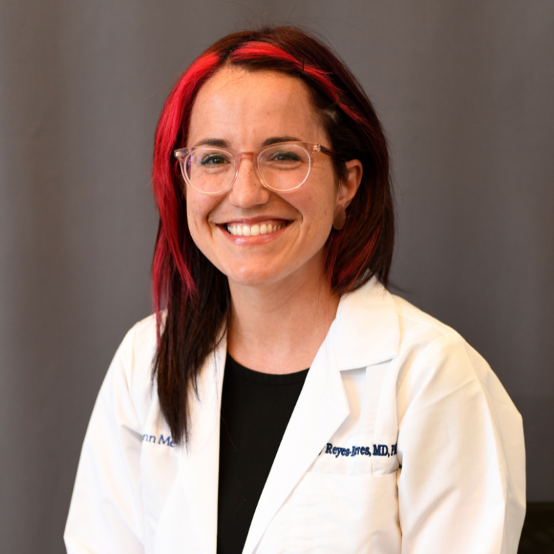 Research Study: Promoting brain hematoma clearance via immunomodulation of infiltrating macrophages.
Primary Investigator: Sahily Reyes-Esteves, MD, PhD, Instructor/Incoming Assistant Professor (CE Track), University of Pennsylvania
Background: Intracranial hemorrhage (ICH) is a deadly form of stroke caused by bleeding in the brain with minimal therapeutic strategies. Among ICH causes, ruptured vascular malformations disproportionately affect younger patients and thus, lead to a significant loss of productive life- years. The recent success of surgical hematoma evacuation trials in ICH suggests that strategies that decreased hematoma burden could be beneficial for patients with ICH, but many are not surgical candidates.
Nanoparticles have been clinically available for decades, including the successful mRNA COVID vaccine packaged in lipid nanoparticles (LNPs). Given LNP’s potential for delivering a variety of cargo to specific cell types, bioengineering of CNS-targeted LNPs could provide powerful therapy for ICH of multiple etiologies, including ruptured vascular malformations. Using a mouse model of ICH, Dr. Reyes-Esteves has developed a vehicle for drugs using tiny drug carriers called lipid nanoparticles (LNPs) to deliver treatment molecules to these macrophages.
Using the ICH model in mice, it was noted that LNPs conjugated to antibodies targeting adhesion molecules (such as the vascular cell adhesion molecule, VCAM) resulted in improved brain drug delivery compared to passive delivery, even at times of high vascular leakage. Based on this, the team built a platform technology for drug delivery to the brain during ICH, which achieved 5x higher brain delivery compared to passive delivery. This proves that reduction in hematoma size is a feasible therapeutic target after ICH that leads to behavioral improvements, and sets the stage for the central hypothesis that LNP-mediated immunomodulation of brain- infiltrating macrophages after ICH can improve hematoma phagocytosis and outcomes in experimental ICH.
Research Objective: The goal of Dr. Reyes-Esteves’ proposal is to develop a new treatment that will use molecular tools to encourage macrophages to remove blood more effectively from the brain while reducing harmful inflammation.
Outcomes: With this grant from the Aneurysm and AVM Foundation, Dr. Reyes-Esteves hopes this research could lead to a non-invasive therapy that helps more ICH patients recover with fewer complications.
Source: Dr. Sahily Reyes-Esteves, MD, PhD. This research summary has been adapted and edited from Dr. Reyes-Esteves’ research proposal.
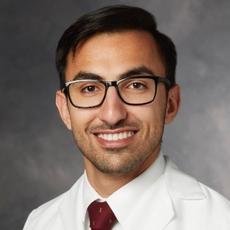 Research Study: Trial of Small Molecule Inhibitors in a Novel, Localized, Inducible, Sporadic Brain Arteriovenous Malformation Mouse Model.
Primary Investigator: Dr. Rashad Jabarkheel, Department of Neurosurgery, Perelman School of Medicine, University of Pennsylvania
Background: As members of the TAAF community well know, the diagnosis of a brain AVM is devastating. Brain AVMs are complex vascular lesions where there is an abnormal tangle of blood vessels such that high flow arterial blood is directly shunted into veins, bypassing a normal capillary bed. Brain AVMs are high risk lesions because the abnormal blood flow within them makes them prone to rupture and consequently cause massive brain bleeds. Treatment of brain AVMs is challenging. For at least a third of brain AVMs, which are high grade, curative microsurgery is not an option, and any currently available treatment option is associated with poor outcomes. Unfortunately, no medications exist to decrease the size of brain AVMs and make them go away.
The goal of Dr. Jabarkheel’s project is to develop an effective medical treatment option for brain AVMs. It has recently been found that the majority of brain AVMs are due to mutations in a specific signaling pathway in the body called the KRAS pathway. Interestingly, the KRAS pathway is also involved in nearly half of all human cancers, and just in the past couple of years new KRAS pathway inhibitors have been developed for this purpose. This proposal wishes to test whether these KRAS pathway inhibitors can not only treat cancer, but also cause brain AVMs to regress.
To do so, Dr. Jabarkheel has developed a mouse model of brain AVMs that allows the team to monitor AVM size continuously. The mice will be injected with AAV-Dre through a cranial window at 6-8 weeks of age. The mice will be monitored for the development of AVMs by visible light imaging at post-operative day and fluorescein angiography for 4 weeks. The primary endpoint of the study is measured frequency of AVM regression.
Research Objective: The goal of Dr. Jabarkheel’s proposal is to trial whether KRAS pathway inhibitors can cause brain AVMs to regress in mice.
Outcomes: Using this grant from the Aneurysm and AVM Foundation, Dr. Jabarkheel hopes the research will set the stage for clinical trials of KRAS pathway inhibitors for treatment of human brain AVMs.
Source: Dr. Rashad Jabarkheel, Department of Neurosurgery. This research summary has been adapted and edited from Dr. Jabarkheel’s research proposal.
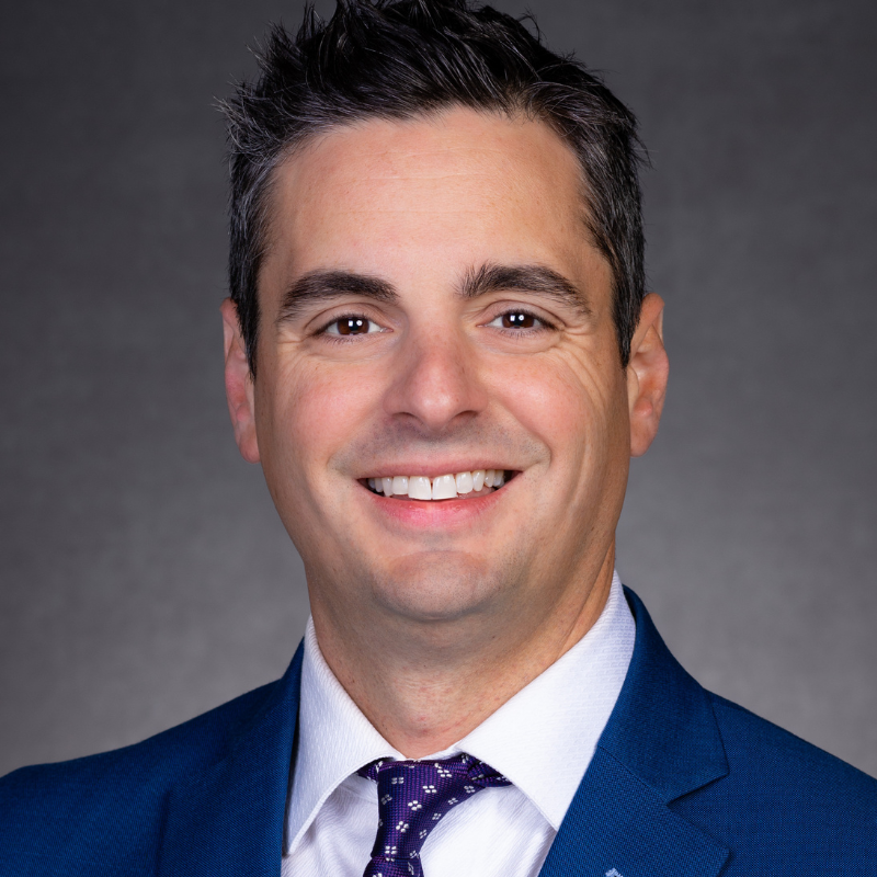 Research Study: Patient-Specific CFD Modeling of Intracranial Aneurysms: Distinguishing Hemodynamics of Aneurysms with and without Blebs with Inlet Boundary Conditions from 4D flow MRI.
Primary Investigator: Isaac Josh Abecassis, Assistant Professor, University of Louisville School of Medicine
Background: Intracranial aneurysms (IAs) can be devastating if they rupture, with almost half the patients dying and the remainder with significant disability. Sometimes IAs develop blebs or daughter sacs, which can indicate a spot of weakness in the aneurysm wall. Blebs can also develop on other cerebrovascular pathologies, like AVM draining veins. While computational fluid dynamics (CFD) can estimate important flow-related features that are associated with rupture, at least to date, most of these models do not factor in patient-specific biological conditions (i.e. how stiff the blood vessel wall is, how viscous the blood is, and the patient’s blood pressure). We aim to determine the difference in flow conditions between aneurysms with blebs from those without blebs.
4D-Flow magnetic resonance imaging (4DF-MRI) is a patient-specific, noninvasive MRI technique that collects detailed blood flow information from patients directly while inside an MRI scanner. This information can be used to inform more accurate CFD models; however, it has substantially lower resolutions. We propose to develop high resolution CFD models which utilize 4DF-MRI in patients with IAs, with an emphasis on the presence of blebs, resulting in highly accurate flow-related parameters (like Wall Shear stress (WSS) and Oscillatory Shear Index (OSI)). Understanding flow conditions within IAs and near blebs will help clinicians triage “higher risk” versus “lower risk” aneurysms.
Ultimately, the results of this investigation will help to develop a real-time “CFD-like” analysis on aneurysms, blebs associated with aneurysms, and AVM draining veins with and without venous varices to determine the risk of bleeding. Future studies will also focus on specific aneurysm locations that are more prone to rupture (i.e. Anterior communicating artery) to determine if these flow- related parameters are different based on location.
Research Objective: The goal of Dr. Abecassis’ proposal is to develop patient-specific CFD models based on 4DF-MRI in patients with IAs with and without blebs, and derive key parameters that have shown prognostic value in predicting aneurysm rupture.
Outcomes: With this grant from the Aneurysm and AVM Foundation, Dr. Abecassis’ hopes this can prompt more studies in the future to determine if these flow- related parameters are different based on location.
Source: Dr. Isaac Josh Abecassis, Assistant Professor. This research summary has been adapted and edited from Dr. Abecassis’ research proposal.
 Research Study: Precision Gene Correction for Sporadic KRAS-Driven Brain Arteriovenous Malformations.
Primary Investigator: Pazhanichamy Kalailingam, PhD, Harvard Medical School
Background: Brain arteriovenous malformations (bAVMs) are high-flow anomalous connections between cerebral arteries and veins that bypass the intervening capillary network, leading to a high risk of intracranial hemorrhage, often causing death or lifelong disability.Sporadic brain AVMs (bAVM), affecting approximately 15 per 100,000 people per year in the US2,, are often caused by somatic mutations, particularly in the KRAS and BRAF genes in > 60% of cases. Treatment options include open neurosurgical resection, endovascular embolization, and stereotactic radiosurgery (SRS) but are often ineffective for large and eloquent lesions.
Hemorrhage of bAVMs can be lethal, with a ~4%15 annual rupture rate (compounded over one’s lifetime, as most bAVM are diagnosed in childhood), and often go undetected until symptomatic. Moreover, residual deficits after hemorrhage include epilepsy, complex headache, cognitive and pain syndromes. Without effective treatments, bAVMs remain “ticking time bombs,” underscoring the urgent need for non-surgical, molecular-targeted therapies.
Somatic gain-of-function mutations in KRAS and BRAF have been identified in 60–81% of sporadic bAVM, are present at low allele frequencies (0.5–4%), and are primarily enriched in endothelial cell (EC) populations. These mutations disrupt EC function, cause abnormal EC size and migration, together driving bAVM formation and predisposition to hemorrhage. Among these mutations, the KRASG12D mutation is the most prevalent (32-52.4% of cases; ~30,000 patients in the USA), making it an ideal target for therapeutic intervention. While small molecule therapies targeting the KRAS pathway, such as those developed for cancer, show high toxicity due to lack of selectivity for mutant KRAS, recent allosteric inhibitors for KRASG12C have shown promise in reducing AVM size (extracranial) in limited cases. These studies demonstrate the feasibility of inhibiting KRAS activity as a viable strategy for bAVM treatment in humans.
Research Objective: The goal of Dr. Kalailingam’s proposal is to optimize CRISPR base editing to correct the KRASG12D mutation in human and mouse endothelial cells, restoring normal function and reducing in vivo AVM formation.
Outcomes: Using this grant from the Aneurysm and AVM Foundation, Dr. Kalailingam hopes this research can lay the foundation for future translational efforts to develop targeted gene therapies for AVM patients, addressing an urgent, unmet clinical need.
Source: Dr. Pazhanichamy Kalailingam Senior Scientist. This research summary has been adapted and edited from Dr. Kalailingam’s research proposal.

 Research Study: A novel tyrosine kinase inhibitor coated flow diverting stent as a drug delivery system for intracranial aneurysms
.jpg) Research Study: Targeting cell adhesion molecules for treating brain arteriovenous malformation
Research Study: Targeting cell adhesion molecules for treating brain arteriovenous malformation Research Study: A Prospective Study Examining Mobility and Sleep after Aneurysmal Subarachnoid Hemorrhage using a Wearable Fitness Tracker
 Research Study: Role of IL-17 in Neurovascular Dysfunction after Subarachnoid Hemorrhage
.jpg) Research Study: Elucidating the genetic basis of Vein of Galen malformation
Research Study: Elucidating the genetic basis of Vein of Galen malformation Research Study: Enhancing glymphatic-lymphatic clearance as a novel strategy to treat SAH
 Research Study: Overcoming barriers in Therapeutics, Imaging and Biomarkers for brain AVMs
 Research Study: Prevention Of Rbpj/Notch Mediated Brain Arteriovenous Malformation Via Blockade Of Apelin Or Osteopontin Signaling In Mice
 Research Study: Piezo1 mechanoreceptor overexpression and dysregulation as a novel mechanism of intracranial aneurysm development
 Research Study: Detection of activating somatic mutations in vascular malformations using minimally-invasive techniques
 Research Study: Targeting KRAS signaling in brain arteriovenous malformations
.jpg) Research Study: Developing and Evaluating an Automated Patient Monitoring System for Preventing Fragmentation of Subarachnoid Hemorrhage Care: A Learning Health System Approach
Research Study: Developing and Evaluating an Automated Patient Monitoring System for Preventing Fragmentation of Subarachnoid Hemorrhage Care: A Learning Health System Approach Research Study: Implementing Auricular Trancutaneous Vagal Nerve Stimulation Following Subarachnoid Hemorrhage To Modulate Neuroinflammation And Improve Outcome
 Research Study: Assessment of RNA Expression of Adhesion Molecules, Mechanoreceptors, and Inflammatory Proteins in Endothelial Cells Harvested From Unruptured Intracranial Aneurysms in Adults.
ThompsonPh.D.Headshot.jpg) Research Study: Evaluating the use of Remote Ischemic Preconditioning in Patients Undergo Endovascular Repair of Brain Aneurysms
Research Study: Evaluating the use of Remote Ischemic Preconditioning in Patients Undergo Endovascular Repair of Brain Aneurysms Research Study: Neutrophil Infiltration in Coiled Aneurysm Healing: Adjuvant IL-17 Blockade
 Research Study: Human Genetics and Molecular Mechanisms of Vein of Galen Malformation
.jpg) Research Study: Patient-specific Genetics of Cerebral Aneurysm Endothelium
Research Study: Patient-specific Genetics of Cerebral Aneurysm Endothelium Research Study: Mechanisms of Neurocognitive Decline After Subarachnoid Hemorrhage
.jpg) Research Study: Histological and Blood Flow Evaluations of AVM and Cerebral Artery Vasculature to Create a Simple Computational Fluid Dynamic Model of Arteriovenous Malformations
Research Study: Histological and Blood Flow Evaluations of AVM and Cerebral Artery Vasculature to Create a Simple Computational Fluid Dynamic Model of Arteriovenous Malformations Research Study: Ceruloplasmin concentration and ferroxidase activity in CSF and risk of brain injury after aSAH
 Research Study: An Investigation of Epoxyeicosatrienoic Acids as a Treatment Strategy to Improve Glymphatic Flow, Cerebral Blood Flow and Behavioral Outcomes Following Subarachnoid Hemorrhage in Rats
 Research Study: Quality of Life in Patients Diagnosed with Unruptured Cerebral Aneurysm: Prospective Single-Center Series